Topical problems of hunter NLS diagnostics,metatron nls parts 5


download url
Introduction
The morphological diagnosis of thyroid gland pathology during surgery
is rightfully considered one of the most important and complicated tasks
faced by anatomist and surgeon. This research aimed to choose an optimum
surgical tactics with tumors and tumor?like lesions of the thyroid gland,
which is achieved by accurate verification of the process as well as by deter?
mining its spread in the organ and/or beyond it. The experience in the use of
nonlinear computer diagnostics (NLS) in surgical clinics covers quite a short
period of time, during which quite conflicting opinions w.r.t. its efficiency
were formed. Among the prime considerations against an extensive use of
NLS we should mention the possibility to preclude in some instances malig?
nant pattern of a new growth because of a morphological similarity of follic?
ular tumors. Without down?grading this problem and based on our own
experience in the use of NLS we have attempted to access the importance of
this method for choosing the optimum surgical tactics and working out some
methodical techniques enhancing efficiency and accuracy of the NLS inves?
tigations.
Subject and investigation methods
We analyzed the results of 682 pathohistological investigations of the
thyroid gland carried out in 2000?2001 in patients operated on for solitary
nodes, diffuse and multinodal hyperplasia and autoimmune thyroid diseases.
326 of all surgeries were accompanied by NLS. NLS data were compared
with the final results of pathohistological investigations.
Result analyses
Of 682 surgical operations of the thyroid gland, 326 (47.8%) were
accompanied by NLS?investigation. According to our information, there is
an increased demand for NLS, which has to do with a growth of surgical
operations for nodular goiter from 70% to 85% and also with a growing thy?
roid cancer incidence including hyperplastic and autoimmune lesions of the
57
MR?signal. Similar nodi were detected in the dorsal part of the pons on the
left and in the basal regions of the frontal lobes. In the frontal lobe the exam?
ination subcortically detected an ellipsoidal cyst 1.2×0.5 cm. In the T?1 pic?
ture of the field the nodi detected in the T?2 picture produced a slightly
diminished MR?signal and was clearly outlined. Upon administration of
magnevist some nodi, not visualized in the T?1 picture, manifested them?
selves by uniform amplification of the MR?signal, the others produced an
amplification in the form of a thin ring or a small amplification in the center.
The cyst did not respond to administration of the contrasting agent. The
central regions of the granulomatous fields represented by necrotic zones
were more hyperintensive in the T?2 picture and after the contrasting agent
administration did not accumulate it, a signal amplification occured in the
peripheral regions in the form of a thin ring. The lateral ventricle bodies were
not quite clearly enlarged. The median structures did not appear to be shift?
ed.
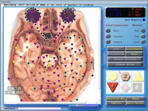
In the frontal lobe on the right NLS picture shows subcortically detect?
ed hyperchromatic areas (6 points) surrounded by a perifocal edema zone (3?
4 points) detected in the dorsal part of the encephalon on the right. A spec?
tral similarity to the standard reference process “toxoplasma gongii” (D <
0.183) was found, which allowed to confirm the diagnosis of toxoplasmosis.
56
tumors of the thyroid gland. A comparison was made considering age and
gender of patients as well as the size of the tumorous nodes in 61 cases of fol?
licular cancer and 162 cases of follicular adenoma. The study of the parame?
ters didn’t detect any difference between these two patient groups. The
male/female ratio in both groups was the same ? 1:9, the average age of the
patients operated on for adenoma was 42.36±13.76 and was not different
from that of the patients in the follicular cancer group (41.40±16.14).
Follicular cancer is known to be more common with elderly people and very
rare with children and teenagers. The latter circumstance could be a supple?
mentary reference point for investigating solitary nodes of the thyroid gland
in junior patients. The investigation analysis of 89 cases of follicular tumors
in patients operated on at the age of 30 showed that in one third of cases the
new growth was of a malignant nature.
Some differences were found in the average diameter of the tumorous
nodes: 3.05±1.45 cm for adenoma and 3.89±1.77 for follicular cancer (p <
0.05). At the same time, the coincident size limits (from 1.5 to 8 cm) in
patients in both groups made this evidence an unreliable indication in the dif?
ferential diagnosis of tumors. Both kinds of neoplasms equally often (approx.
in 80% of cases) were not accompanied by morphologically significant
changes in the thyroid gland being a solitary node. Some frank secondary
changes such as sclerosis, petrifaction, cystic changes, hemorrhages, e.t.c,
were more often observed in follicular adenomas, however these distinctions
were not authentic enough.
In our opinion the difficulties in the clinic morphological interpretation
of the follicular tumors pattern need more than anything else improvement in
the methodical techniques, which is especially important considering certain
time and hardware?related limitations of the NLS?method. When faced with
diagnostic difficulties we investigate series of nidi in every 30?60 mm.
Conducting investigation on such a large scale consumes additional time (30?
45 min) and yet in many instances it allows to specify the pattern of a follic?
ular tumor. If the investigation of some additional nidi doesn’t produce the
desirable result, the diagnostics is performed after the surgery. According to
our observations, in 65% delayed cases the tumor proved to be malignant yet
had minimal manifestations of invasive growth into the node capsule or its
individual vessels. Absence of palindromium in 95?99% of cases, following
the surgery for follicular cancer with minimal manifestations of invasive
growth into the capsule and with some individual vessels (up to 5) involved,
gives solid grounds to classify these tumors as clinical “boundary” processes
whose malignant potential remains conditional and justifies the tissue?spar?
ing amount of the thyroid resection (lobectomy with isthmectomy and subto?
59
thyroid gland. We did not succed in specification of the pattern of the process
in the course of 3.6% of all the surgical operations. In 65% of cases for which
diagnosis was postponed, a malignant process was detected in the final phase
of the investigation. An erroneous intraoperative diagnosis was made in 4.8%
of cases with hyperdiagnosis of thyroid cancer recorded in 5 cases.
Carcinoma was not identified during 38 operations; in 23 cases of the surgery
the tumor did not exceed 2.5 cm nor did it spread beyond the thyroid gland.
17 (2.3%) patients needed correction of the amount of the thyroid gland
resection, which was done on the 4?5th day after the first surgery. The NLS
sensitivity was 76.4%, specificity? 87.6% and accuracy? 78.6%.
The submitted data generalizing the experience in the use of NLS in a
specialized surgical clinic are indicative of extensive opportunities for choos?
ing the optimum surgical tactics to treat the goiter using this method, and
also of a growing recognition of NLS despite some recent publications ques?
tioning the efficiency of consultations during the surgery. Apart from some
evidence of a high efficiency rate, another advantage of NLS method is indis?
putably the low percentage of delayed diagnoses, which in our cases was
under 1.4%. The advantages of the method include quickness (10 or 15 min)
and relative technical simplicity of the investigation.
According to our research and some literary evidence, the problems in
the course of NLS?investigation are caused by differential diagnostics
between cellular follicular adenomas and minimum invasion follicular can?
cer. These very cases account for the major share of delayed and erroneous
results. In the series of our investigations in 48 cases of tumors with a micro
follicular or trabecular structure (among which 21 were benign and 27 malig?
nant) drawing a final conclusion on their pattern was impossile. In 5 cases it
was false negative. At the same time 257 follicular adenomas and 30 cases of
follicular cancer were correctly verified in the course of surgery.
It is known that the problems of differential diagnostics of follicular ade?
noma and follicular cancer are closely related to histotypical and cytotypical
similarity of the two processes, that are so much expressed that it is impossi?
ble to diagnose a carcinoma without apparent manifestations of a malignant
potential in the form of the tumor ? germinated capsule with the tumorous
invasion into its vessels. With cancer having minimum invasion the nidi of
infiltrative growth appear to be isolated. In addition, being microscopically
invisible these diagnostically important areas may escape observation in case
of a limited number and random choice of investigation targets in the course
of surgery.
We have considered a number of clinical and microscopic characteristics
in terms of their potential use in NLS differential diagnosis of follicular
58
obtained from other clinics, a tumor in excess of 0.5 cm incurs a higher risk
of potential palidromium and requires a more radical approach to its treat?
ment, than a microscopic cancer nidus. In this connection, it is recom?
mended to take a series of parallel shots of the thyroid tissue in different pro?
jections in the course of macroscopic search for cancerous nidi. According to
our observations this technique may be effective in diagnosing 52% of papil?
lary micro carcinomas sized up to 0.4 cm and 68.6% of tumors over 0.4 cm,
and in most cases will ensure the right choice of a surgical approach.
According to most pathologists, diagnostics of follicular version of pap?
illary cancer is considered to be one of the most difficult problems in NLS?
investigations. Erroneous verification or this neoplasm often leads to anoth?
er surgical operation. Among the difficulties in diagnosing this kind of tumor,
we should first mention some artificial histogram changes in tumors cells,
which hamper identifying diagnostically significant cytological criteria of
papillary cancer. In our series of observations follicular version of papillary
cancer was reported in 43 cases, in 2 of which diagnosing was postponed until
after final investigations and in 5 (3.2%) cases the diagnosis was false nega?
tive. None of the cases showed any metastatic lesions of cervical lymph
nodes. Considering the spectral similarity of follicular encapsulated version
of papillary cancer to adenomatous goiter, the cytological differences in dif?
ferential diagnosis are the decisive criterion in differential diagnosticsis. In
order to assess them in doubtful cases we have additionally investigated some
impression smears, which in most cases helped detect some changes in the
nuclei characteristics of papillary thyroid cancer, such as irregular shape,
jagged boundaries, deep nucleolemma invaginations, outlines of intranuclear
sulci and inclusions marked off by marginally condensed chromatin, fine
chromatin dispersion, etc., as well as to indirectly assess some cohesive prop?
erties of tumorous cells and the inflammatory infiltrate pattern. Among some
cancer?suspicious histological signs observed on a frozen section, we can
note polymorphism of follicles lined with high cubical epithelium with
intensely tinged colloid (if fixed in ethanol!) and/or its marginal vacuolation,
close adherence of follicles to one another owing to scarce stroma in the cen?
tral part of the node, hemorrhage in the follicle lumen at abundance of
siderophages, multinuclear cells, etc.
The recent years publications have extensively debated expediency of
NLS?investigations of the thyroid gland in the cases with the available results
of aspiration biopsy. The cytological investigation in known to be the most
extensively applicable method of presurgical diagnostics of nodular forms of
the goiter because of its accessibility, comparatively low cost, lack of trauma?
tism and most importantly, high accuracy. The progress in the diagnostics of
61
tal thyroidectomy) similar to the one recommended for surgery for follicular
adenoma.
Thus, the NLS?investigation of follicular thyroid tumors can be regard?
ed as an efficient method for choosing the optimum surgical approach,
because with sufficient experience and proper performance it allows to diag?
nose some clinically adverse forms of follicular thyroid cancer that actually
need radical surgery and post surgical treatment.
The papillary cancer is the most common form of thyroid gland carci?
noma. It was correctly diagnosed by means of the NLS?methods in 63.2% of
cases, was a diagnosis failure in 26.3% and was responsible for delayed diag?
nosis in 0.6% of the observations. Unlike follicular tumors, most versions of
papillary cancer typically have frank histotypical differences from benign
proliferate processes, clear manifestations of infiltrative germination in the
tumor?surrounding tissues and frequent metastatic lesion of the lymph nodes
by the time of surgery, which allows to diagnose a malignant process without
difficulty even with inadequate practical experience. The difficulties we con?
fronted mostly concerned papillary micro carcinoma, which made 71% of
cases nondiagnosed during the surgery, and also a follicular encapsulated ver?
sion of papillary cancer that was responsible for the rest 29% of diagnostical
errors.
The problem of NLS papillary micro carcinoma lies in a macroscopic
search for a cancer nidus in the removed fragment of the thyroid gland, which
creates difficulties because of concomitant changes in the thyroid gland at a
multinodal form of the goiter or autoimmune processes. At the same time in
45 cases microcarcina was diagnosed intraoperatively, including 15 cases,
where the tumor size was below 0.4 cm. In 23 cases of papillary micro carci?
noma unidentified by NLS, the surgery was performed for multinodal goiter
(15 cases) and autoimmune thyroiditis (8 cases), which determined the
required amount of surgical intervention (subtotal or total thyrodectomy).
The undiagnosed micro cancer nidi sized from 0.4 to 0.9 cm did not spread
beyond the thyroid gland and afterwards none of the patients affected by
latent carcinoma required another surgical operation to extend the amount of
thyroid gland resection.
Papillary micro carcinoma is known to localize quite often in the thyroid
gland, especially in elderly people and it does not always display its malignant
potential in the form of clinical implications (I.L. Avetisyan, 1999). The
progress of the great majority of such tumors is entirely favorable.
Meanwhile, a direct relationship was established between the size of a papil?
lary micro carcinoma nidus and the frequency of its metastasizing into the
cervical lymph nodes. According to our information and some evidence
60
From comparion of the FNAB and NLS results it was found that of 27
false negative results of punch biopsy 21 nidi of malignant growth were
detected during surgery among multiple benign goiter nodes. The latter,
being prevalent in clinical implication, became the object of FNAB leaving
some latent cancer nidi sized 0.2?1 cm undiagnosed before the surgery. The
insufficiently accurate cytological interpretation of the pattern of a cystic
cavity in the 111G also requires an intraoperative verification. In our series in
2 cases of encapsulated papillary cancer with cystic generation wrongly inter?
preted as a benign process during FNAB the diagnosis was rectified by means
of NLS.
Another equally important task of intraoperative investigation is leveling
possible false positive conclusions of FNAB. In our series in 497 patients hav?
ing benign new growths according to FNAB results, papillary cancer was sus?
pected in 2 cases and a malignant process was not excluded in 26 cases. The
rectification of the process pattern in the course of surgery allowed to choose
the most efficient surgical approach in all cases.
Conclusions
1. The NLS?investigation of thyroid tumors is an efficient method for
choosing surgical approach in surgery for nodal and diffuse forms of goiter.
2. The NLS performed for the patients following FNAB, considerably
enhances the accuracy of morphological investigations at the preliminary
phase, and optimizes the surgical approach in surgery for the thyroid gland.
3. This is a very important reason in favour of the appropriatness of using
NLS?investigation together with FNAB.
63
thyroid tumors using the cytological investigation technique has induced
some researchers to become result?oriented in choosing a surgical approach.
In this connection we made an attempt to define to what extent this
approach is justified after having analysed our own observations and the prac?
tical experience of specialists in a number of well?known clinics of the world.
Among some major problems of fine needle aspiration biopsy (FNAB)
performed under the control of ultrasound scanning (US) we can mention an
amount of aspirate inadequate for diagnosis as well as some situations that do
not allow to preclude a malignant process in the node under the investigation.
While in the former instance a repeated procedure may prove efficient for
30% of the patients, the other problem needs to be solved by a surgical
removal of the tumor with a subsequent histological verification of its pattern.
According to some publications, the number of tumors, which malignant
potential cannot be excluded based on FNAB results, exceeds 11%. In the
majority of cases (about 70%) this kind of diagnostic problems is caused by
follicular adenoma.
According to our information of 338 patients who had surgery after pre?
liminary FNAB the malignant process was not cytologicaly ruled out in 41
cases. In 26 of these observations follicular adenoma and in 15 thyroid can?
cer were verified (2 follicular, 3 medullary and 10 papillary carcinomas). In
all cases the NLS allowed to specify the diagnosis and avoid errors in defin?
ing the amount of resection.
The problem of FNAB accuracy is no less pertinent. According to some
recent reports, the sensitivity and specificity of thyroid FNAB has approached
100%. At the same time, it proves to be difficult to interpret the obtained data
due to different approaches to their analysis. For instance, specialists in some
clinics, where the results were highly accurate, consider only specific cytolog?
ical conclusions ignoring a category of tumors, which cytological picture gives
grounds only to suspect malignant change. In analyzing the FNAB accuracy
some researches consider follicular adenoma in the same category as malig?
nant tumors. Though this kind of approach may be justified in terms of indi?
cations for surgery, by no means it can be justified in terms of a surgical
approach to be chosen. The analysis of some publications shows, that FNAB
can ensure the right surgical approach only in 70?75% of cases. The FNAB
efficiency data evaluated after classifying cancer?suspicius conclusions as
malignant tumors and adenoma as a benign tumor, were as follows: sensitivi?
ty ? 92.1%, specificity ? 94.4%, accuracy ? 93.45. Thus, the FNAB data could
help decide on the proper amount of surgery for 90% of the patients, which in
terms of specialized clinic cannot be a sufficient reason and is an argument in
favour of supplementing FNAB with NLS?investigation.
62
up new opportunities for detecting diseases in the hepatopancreatoduodenal
region, with obstructive jaundice being one of their main clinical implica?
tions. With the development and adoption of a number of speedy programs
for obtaining NLS images, specifically NLS? cholangiopancreatography,
which allows to obtain an integrated virtual picture of the biliary system and
pancreatic ducts without administration of contrasting agents and interven?
tion into the biliary system, the method was attempted to put into active use
as an alternative to ERCPG.
Some published works dealing with NLS have some distinct trends to
pay more attention to this issue with a view of obtaining sufficiently convinc?
ing informaion, that would allow to draw a final conclusion about a new rela?
tionship between integrated X?rayendoscopic examinations, and in the first
place between ERCPG and NLS, when detecting a pathology in the
hepatopancreatoduodenal region. Some of the works suggested that NLS be
used as a method preceding endoscopic cholecystoectomy.
With all the above in view, this paper aims to present our data on the role and
significance of NLS at certain diseases in the hepatopancreatoduodenal region.
To achieve this aim the following tasks were performed:
1. examination of the test group to study different versions of a standard
NLS?picture of the biliary tract;
2. description of the principal NLS semiotics in the patients with a
pathology in the hepatopancreatoduodenal region;
3. cross?comparison of MRT, ERCPG and NLS for a more objective
assessment of the collected data;
4. definition of clinical indications and diagnostic potentials of the NLS
method for the patients with obstructed biliary ducts.
Subject and methods
The NLS investigation was performed on 54 patients, of them 19 made a
test group and 35 had different pathologies in the hepatopancreatoduodenal
region, with 89% of these patients showing signs of obstructive jaundice. The
patients were from 36 to 77 years old. There were 20 women and 15 men in the
group of 35. As a primary method of investigation all the patients had a sonog?
raphy which acted as a screening tool for performing NLS. A relative com?
parison of the results of MRT, ERCPG and NLS was made for 18 patients.
The NLS investigation was carried out using the “Oberon” metatron
jointly manufactured by the Institute of Practical Psychophysics and Clinic
Tech Inc. (USA) and equipped with a 4.9 Ghz trigger sensor.
We assessed the condition of the lymph nodes, especially in the portal
fissure projection, and the hepaticoduodenal ligament on the virtual images.
65
Nonlinear computer diagnostics
and the problem of pathology
in the hepatopancreatoduodenal area
S.P. Tokar, A.S. Davydova,
T.L. Guseva, V.I. Gusarov,
Z.F. Khabibullina, L.S. Pugacheva
The problem of pathology in the hepatopancreatoduodenal area still
remains urgent and explains why researches are keen on the search for
improved diagnosis methods, since the diagnostics proper is the starting point
for determining an approach to treatment. Today the basic methods for diag?
nosing a pathology in this region are traditionally methods of direct artificial
contrasting of the pancreatobiliary system, such as endoscopic retrograde
cholangiopancreatography (ERCPG) and percutaneous transhepatic
cholangiography (PTCG), well established in both diagnostics and treatment
of a number of diseases such as cholelithiasis, cysts and tumors in the head of
pancreas, tumorous and corrosive strictures of the biliary ducts, tumors of
Vater’s papilla, etc.
At the same time, the radioendoscopic methods of investigation of the
biliary ducts, though characterized by a rich diagnostic informational con?
tent owing to their invasiveness, still do not eliminate the danger of serious
complications, such as acute pancreatitis, hyperamylasemia, cholangitis,
sepsis, and allergic reactions, biliary flux into the abdominal cavity with
developing biliary peritonitis, hemorrhages, ect.
Their incidence rate varies from 0.8 to 36%. Besides, in the course of
ERCPG different technical problems may arise (failure in the cannulation of
Vater’s papilla, the impossibility to enter the duodenoscope at esophagus dis?
eases, such as strictures, achalasia, ect.). In addition ERCPG requires
involvement of certain specialists like radiologic diagnosticians, surgical
endoscopists and anesthelists.
The advent of new diagnostical techniques in radiology and first of all
ultrasound scanning (US) and computer tomography (CT), did not produce
a great limiting impact on the use of ERCPG so far as these methods were not
successful in solving a number of diagnostical issues related to pathologies in
the biliary system and pancreas. The development of nonlinear computer
diagnostics (NLS) as s method for diagnosing abdominal pathology, opened
64
The major NLS disadvantages in diagnosing concrements in the hepatic?
ocholledochus are associated with certain difficulties in assessing the chole?
dochus condition, when the choledochus is fully filled with concrements.
In 1 observation the concrement localized in some distal areas of the
hepaticocholledochus, and on NLS shots it looked like a hyperchromogenic
oval?shaped defect with the upper outline looking like a concave lens. The
combination of NLS?shots with conventional MR?tomograms in axial plane
allowed us to specify the spatial relationship between the choledochus and head
of pancreas and the duodenum, in other words, it allows to detail the localiza?
tion of the concrement in the ampullar region of the common biliary duct.
Papillosphincterotomy was done during ERCPG with concrement
extraction.
The genesis of benign strictures of biliary ducts was related to their sur?
gicial lesion or inflammation caused by lithiasis, chronic pancreatitis or
papillostenosis in 90?95% of cases. The number of iatrogenic lesions of the
biliary system ducts grew up with the extensive application of the laparo?
scopic cholecystectomy, because the intraoperative investigation of the com?
mon bile duct is more complicated during laparoscopy than during open sur?
gery. In this connection, in terms of preoperative preparation for endoscopic
cholecystectomy it is necessary to specify the anatomy of the pancreatobiliary
system and assess its condition in order to prevent potential iatrogenic lesions
of the biliary ducts.
So, owing to its noninvasiveness and high resolution, NLS can be a diag?
nosis?determining method for this kind of patients. Unlike ERCPG, NLS
allows to visualize the bile ducts above and below the obstruction level, which
is displayed on both MRT and NLS shots. The latter method gives a virtual
physiological picture of the condition of hepatic and pancreatic ducts as
compared to ERCPG, in which the administration of a contrasting agent
overstates the extent of duct dilatation.
In all of our observation NLS allowed to define the accurate extent of
the arctation, its length and cause. In 2 cases the arctations localized at the
cystic duct level, which was indicative of their iatrogenic genesis. In 1 case it
was an arctation hepaticocunoanastomosis. In 5 observations the arctations
from 1.5 to 2.0 cm long were located at the confluence and in the proximal
region of the hepaticocholledochus.
In assessing the arctation extent in the case where the lumen was not
visualized on NLS?shots, we always analyzed the native MRT scans and sup?
plemented the investigation with thin sections, which allowed forming a
more exact opinion about the arctation extent. At the same time, comparing
the results of nonlinear diagnosis to ERCPG one must admit that the latter
67
We used the “Metapathia IT”, a special computer program for acquisition of
a virtual image of the biliary system and Wirsung’s duct.
Analysis of results
The virtual model distinctly visualized the common bile duct, common
hepatic duct, right and left lobar ducts and gall bladder (GB). The segmen?
tary and subsegmentary intrahepatic ducts are not actually visualized even in
a polyprojection examination. The normal lumen of the common bile duct is
0.6 cm; the NLS?signal coming from it, is homogeneously normochromatic
(1?2 points according to Fandler’s chromatic scale).
The anatomical variations and abnormal developments occur very sel?
dom, yet we observed 3 cases of this kind, of them 2 contained an abnormal
drainage of the cystic duct and 1 an atypically high point of entry of the cys?
tic duct into the common hepatic duct. An insufficient detailing of the papil?
lo?sphincter region is the basic limitation of NLS in our investigation was.
Calculi are known to be the most frequent cause of the bile duct obstruction.
According to our investigations, cholecysto?choledocholithiasis made 34% of all
diagnosed pathologies in the hepatopancreatoduodenal region. Regardless of
their location, the concrements in the biliary ducts were visualized on the NLS
images as individual or multiple hyperchromogenic zones (5?6 points), rounded
or oval?shaped. The sizes of the concrements detected in the hepaticocholle?
dochus and lobar hepatic ducts varied from 5 to 20 mm. 6 patients had single
concrements, and 4 had multiple concrements, and the entire lumen of the
hepaticocholledochus “stuffed” with concrements was found in 1 patient.
The localization of the concrements was variable. In 2 observations the
concrements only localized in the gall bladder and in 5 cases they did in the
hepaticocholledochus; in 1 case the clinic laboratory evidence of obstructive
jaundice was not found, and in 2 cases the concrements were visualized in
both the choledochus and lobal biliary ducts. In 5 cases we observed a con?
currence of concrements in the gall bladder and choledochus.
In the course of our observations we arrived at a conclusion that the NLS?
diagnostics of concrements in the gall bladder depended on their size. So, as
compared to the US data, the concrements under 5 mm in diameter were
largely not visualized on NLS shots, because the signal from them was over?
lapped by a hyperchromogenic signal from the mucous membrane. Small mul?
tiple concrements in the gall bladder that produce a low entropy density signal
(3?4 points according to Fandler’s scale) on NLS?shots hamper their differen?
tial diagnosis because of sediment and putty?like bile. We agree with same
authors who consider the ultrasound scanning to be the “golden standard” in
detecting gall bladder concrements, which should not be replaced by NLS.
66
We observed 4 cases of cholangiocarcinoma with obturation of intra? or
extrahepatic ducts including 2 cases with a tumor localized at the common
hepatic duct level and 2 cases with affected intrahepatic ducts and liver parenchy?
ma. In all of the 4 cases the NLS allowed to precisely localize the lesion level and
define its length. Both cases of cholangiocarcinoma displayed a spectral similar?
ity to the reference standard process ‘liver carcinoma’ (D from 213 to 418).
Researchers observed an increased chromogenic density (5?6 points) of
intrahepatic ducts more proximal to the arctation. In one of the observation,
in the projection of the constriction of the common biliary duct, the MRT
scans displayed a soft tissue structure, up to 3 cm in diameter with a medium
intensity signal, which enveloped the duct in a sleeve?like manner at the
lesion level and was indicative of a tumorous etiology of the structure.
Another observation at stenosis of the common biliary duct with no imaging
of the tumorous tissue, detected enlarged lymph nodes in the lesser omentum
region and a single metastasis into the liver which allowed us to correctly
interpret the pattern of the lesion confirmed by histological investigation of
the biopsy material acquired during transhepatic drainage for decompressing
the biliary ducts. In 1 of the 2 observations of cholangiocarcinoma of intra?
hepatic ducts also histologically confirmed later, we drew an erroneous con?
clusion, because the pattern of MR?changes, i.e., a small ectasia of the intra?
hepatic ducts by a varicose type above the moderately constricted common
hepatic duct, and unaffected hepatic duct more distal from the structure, a
rather long anamnesis of the disease (the patient had had itching fits, occa?
sionally icteric integument, decolored feces and dark urine for ten years) and
the obscure clinical presentation were interpreted by us (and during
ERCPG) as manifestations of a primary sclerogenic cholangitis.
In all three our observations of pancreas head cancer the obstruction of
the biliary duct looked on NLS shots like a progressively growing chro?
mogenic density of the ecstatic biliary duct at the level of its intrapancreatic
area. A frank hyperchromogenic pattern of the intrahepatic ducts was con?
currently noted. The Wirsung duct was unevenly hyperchromatic too. The
standard AUTO TUNE shots allowed to evaluate the spread of the tumor to
the adjacent structures and determine some hematogenic and lymphogenous
metastases. The structure of the tumor itself could be better visualized on the
front shots. In one of the cases we also detected both metastases into the liver,
and enlarged lymph nodes in the suprapancreatic cellular tissue. The histo?
logical investigation confirmed the adencarcinoma in all three cases.
Differential diagnostics of pancreas head cancer and chronic pseudotu?
morous pancreatitis is a very complicated task and until now has been a prob?
lem yet to be solved. Integrated abdominal NLS?investigation with visual
69
method is more exact in determining the extent of duct affection.
However the essential criterion in deciding on the surgical correction
method allows not only to detect the level and length of a structure, but also clear?
ly specify the pattern of cholledochus deformation in presence of an arctation,
which also determines the surgical approach to the reconstructive operations.
The combination of conventional MRT and NLS considerably
enhances the diagnostic potential of this method as oppposed to ERCPG in
diagnosing chronic or acute pancreatitis, because it allows not only to inves?
tigate the condition of the ducts of the pancreatobiliary system, but also to
assess both the pancreas proper and the adjacent organs and structures. Of 4
our observations of chronic pancreatitis in 1 case we had a frank contraction
of the intrapancreatic part of the cholledochus caused by a chronic inflam?
matory process, in 3 patients the constriction of the distal part of the cholle?
dochus was caused by a cyst in the head of pancreas. On the NLS?shots the
obstruction of the biliary duct looked cone?shaped, and its affected part
could be visualized all the way along the head of the pancreas including
Vater’s papilla area, and was assessed at 5?6 points according to Flandler’s
chromatic scale.
In all the cases a moderate chromogenic density of dilated biliary ducts
and a heterochromous response of Wirsung duct occured. The cysts, where
existed, were depicted on MRT and NLS shots and the constricted area of
the common biliary duct had an arc?shaped route because of being forced
back by the cyst. The NLS allowed to detect a relation between the Wirsung
duct and pancreatic cysts.
The most common and well?known causes of the biliary tract obstruction
are the tumors localized in different organs: liver, biliary extra hepatic ducts,
head of pancreas, major duodenal papilla, as well as metastases into the hepa?
toduodenal ligament and portal fissure. In diagnostical department of the
clinical medicine the tumors concentrated in these locations are convention?
ally called the “tumors of the hepatopancreatoduodenal region”. The reason
for that is common clinical implications related to the obstruction of biliary
and pancreatic ducts. In patients affected by malignant tumors localized in
this area the primary symptom of the disease is generally obstructive jaundice.
So, decision on the expediency of surgery for this kind of patients necessitates
an assessment of a clinical prognosis depending on the tumor state according
to the TNM system. So, if a malignant pattern of obstructive jaundice was sus?
pected, then together with the elimination analysis, which is a special pro?
gram, we always carried out standard investigations in the AUTO TUNE
mode which allowed to localize the tumor and assess its spread to the adjacent
structures as well as to define hematogenic and lymphogenous metastases.
68
MECT and NLS in diagnosing
myocarditis of mild or medium gravity
P.S. Bortshov, K.L. Fadin,
O.P. Derkatch, P.A. Abdulov,
T.N. Timofeyefa, B.M. Nikolaev
Introduction
The diagnosis of non?reumatic myocarditis remains a complicated and
pertinent problem, which is conditioned by lack of pathognomonic clinical
signs and similarity of the semiotics of the disease to other kinds of cardiac
pathology.
The notion of myocarditis brings together inflammatory myocardium
conditions, different in terms of etiology and pathogenesis both at isolated
affects of myocardium (primary myocarditis) and at some infectious diseases
and systemic pathological processes (secondary myocarditis). The disease
may develop with a focal or diffuse lesion of the myocardium.
The clinical diagnostics of primary myocarditis presents great difficul?
ties, so because of the lack of direct signs of the inflammatory lesion of the
myocardium, the diagnosis is based on indirect clinical hardware?bsed syn?
dromes that become apparent within 2 or 3 weeks after the administered
infection. The related syndromes are as follows:
1. any newly detected pathological changes in ECG;
2. an increased level of cardio selective enzymes and proteins in blood
(troponin T and I);
3. cardiomegaly as per radiography or echocardiography data;
4. congestive heart failure;
5. changed immunologic indexes (a higher CD4/CD8 and circulating
immune complex ratio, “yes” response to inhibition of lymphocyte migra?
tion;
6. tachycardia, reduced first sound and gallop rhythm.
The needle biopsy of the myocardium is one of such diagnostic tech?
niques. Meanwhile, the basic morphological sign of the disease in this con?
text is presence of an inflammatory reaction (lymphocytic and histiocytic
infiltration) in the myocardium with damaged cardiomyocytes and capillary
endothelium. However, the endomyocardiac biopsy does not ensure an
exhaustive diagnosis because of a chance to pick up a sample from the unaf?
fected section of the myocardium and obtain a false negative result.
71
simulation of changes in the head of pancreas (not always pathognomonic for
a tumor) and its spectral similarity to blastic process (D< 0.425) allowed us
to produce a more definite opinion about the tumorous pattern of the
process. In addition, the value of the NLS consists in its capability to asses
the spreading of the tumour to the cystic duct which is very essential for mak?
ing a decision on the expediency of cholecystodigestive anastomosis.
According to some literary evidence, differential diagnostics of diseases
of the papillosphincteral region by means of NLS investigation is very diffi?
cult. In 1 of our observations of a patient with clinical lab manifestations of
obstructive jaundice the US detected a distal block of cholledochus with
dilated superjacent regions of the biliary system. The duodenoscopy detected
signs of frank papillate of the major duodenal papilla and suspected infiltra?
tion of the longitudinal fold of the duodenum. The percutaneous transhepat?
ic cholangiography (PTCG), that was performed additionally showed a pic?
ture of stenosed papillosphincteral region. The NLS displayed
hyperchromatic intrahepatic ducts and hepaticocholedochus, with the latter
traceable as far as the level of entering the duodenum. The NLS investigation
performed in a sagittal projection allowed to detect an additional structure
with a moderately chromogenic signal in the ampullar region of the cholle?
dochus. It allowed a supposition about a tumorous lesion of the major duo?
denal papilla. The patient died of DVS syndrome. In section: poorly differ?
entiated adenocarcinoma of the major duodenal papilla. We would like to use
this example to stress the importance of a correct methodic approach for car?
rying out an investigation in a specific diagnostic situation.
Thus, our results were able to show that NLS had great prospects for
diagnosing a number of diseases with such a common anatomic?functional
concept as a pathology in the hepatopancreatoduodenal region. So, thanks to
its noninvasiveness, no need for administering contrasting agents and inter?
vening into the biliary system as well as the aibility to combine with conven?
tional MRT, which substantially increases the diagnostic importance of the
method, in quite a lot of cases the NLS investigation can replace radioendo?
scopic methods of investigation (ERCPG and PTCG) as a primary diagnos?
tics method although the other methods can retain their therapeutic func?
tions. NLS may also become a method of diagnostics choice for patients who
have some counter indications to X?rayendoscopic investigation. We believe
it necessary to actively expand the use of NLS with a view of making an ulti?
mate decision on its clinical diagnostic importance.
70
underwent an NLS investigation of the heart within 2?8 months after the pri?
mary radioisotope investigation and three patients had MECT performed
repeatedly 1.5?8 months later.
Depending on the variant of the primary accumulation of leukocytes in
the myocardium, all the patients with clinic?lab signs of myocarditis were
divided into three groups. The first group included patients with a diffuse
accumulation of tagged leukocytes in the myocardium (n=16), the second
one comprised patients with a focal accumulation (n=20) and the third one
did not have any distinct accumulation of tagged autoleukocytes in the
myocardium (n=13). The groups were comparable by age and gender. The
average age of patients in the group with a diffuse accumulation of leukocytes
was 32.1 ± 1.8, in the group with a focal accumulation? 35.2±2.1 and in the
group without a distinct accumulation ? 36.6±2.2.
Result analysis
The analysis of intracardiac distribution of tagged auto leukocytes
showed that all the patients in the first group had a diffuse lesion of the car?
diac walls in at least two regions: the atril 94% of cases (n=15), left ventricle
(LV) 63% (n=10), right ventricle (RV) 63% (n=10) and interventricular par?
tition 63% (n=10). In patients of the second group with a focal accumulation
of auto leukocytes in the myocardium a lesion of the interventricular parti?
tion (IVP) was visualized most often ? in 65% of cases (n=15), and in the
other regions of the heart inflammatory infiltration was detected with cer?
tainty less often than in the first group and was less frank (atrium ? 30%, n=6,
RV?25%, n=5 and LV?20%, n=4).
One of the factors possibly related to the pattern of auto leukocyte accu?
mulation in the myocardium was an extracardiac spot of leukocyte fixation in
the nasopharynx, gall bladder and uterine appendages regions, which could
be regarded as an obscure foci of chronic infection. It was established that at
a diffuse pattern of auto leukocyte accumulation in the myocardium (which
could be interpreted as a sign of diffuse myocarditis) the extracardiac foci of
the infection were visualized in 100% of cases.
With a focal pattern of leukocyte accumulation in the myocardium the
foci of chronic infection were determined in 70% of cases. In the group of
patients without a clear accumulation of the leukocytes in the myocardium
the foci of chronic infections were only detected in 40% of cases. Extra car?
diac foci of tagged auto leukocyte fixation were more often detected in the
nasopharynx, however there were no authentic distinctions among the
groups. Another factor related to the pattern of the auto leukocyte distribu?
tion in the myocardium was the duration of the disease which consecutively
73
According to most authors, the endomyocardiac biopsy confirms the clinical
diagnosis only in 17?37% of cases.
In this connection the development and extensive use in clinical prac?
tice of noninvasive methods of diagnosis of myocarditis that allow to visual?
ize myocardium inflammation and determine its intensity and evolution in
the course of treatment, arouse active interest. We have accumulated some
experience in non?invasive diagnostics of myocarditis and dynamic control
of the myocardium condition in the course of treatment. In the integrated
diagnostics of inflammatory cardiac lesion, additionally to conventional
methods of clinical, laboratory, immunologic and ultrasound investigation,
we have used the methods of monophoton emission computer tomography of
the myocardium (OECT, tomoscintigraphy of the myocardium) with
autoleukocytes tagged by ottTc?hexamethylpropyleneaminooxym
(HMPAO), and NLS?investigations of the heart.
Material and methods
The tomoscintigraphy of the myocardium was performed on the
monophoton emission computer tomograph ‘Elcinct?Apex?SP?6’ (Israel)
every 1?2 and 24 hours after the autoleukocytes reentered the bloodstream.
The leukocytic suspension was extracted from 500ml of the whole blood by
centrifugation and leukocytopheresis. The leukocyte count was 37.000?
45000 cells per 1 microliter. Before its administration the leukosuspension
was incubated in a flask with 99 tTC ?HMPAO (500) for 15 minutes at room
temperature. For better visualization of myocardium inflammation and a
perfusion study the tomoscintigraphy was performed once again using 44tTc?
tetraphosmine. During processing of the results the tomographic ‘sections’ of
the heart were drawn along the short axis every 0.8 cm from top to bottom
and the outward outlines of the aortic ventricle were encircled. Meanwhile
the inflammation areas were visualized on the scans as foci of hyperfixation
of tagged leukocytes in at least three adjacent sections.
The NLS?investigation was carried out using ‘Oberon?4099′ device
equipped with 1.4 GHz digital trigger sensor (IPP ?CLINIC TECH inc). In
analyzing the obtained virtual shots a special attention was paid to the emerg?
ing zones of the increased chromogenic value of the signal from the
myocardium (5?6 points according to Flandler’s scale). All in all 49 patients
affected by myocarditis of mild or medium gravity were examined (25 women
and 24 men aged 15?47). All the patients underwent a primary clinical hard?
ware?based investigation including MECT with tagged leukocytes. 9 patients
had check examinations of the myocardium condition carried out during the
therapy using tomoscintigraphy of the myocardium and NLS. Six patients
72
mulation had NLS carried out 6 and 8 months after MECT, and the shots did
not display any pathological changes.
Thus, the repeated investigations using tomoscintigraphy of the
myocardium and NLS proved, that a clinical improvement in the patients’
condition that could be seen 1.5?3 months after the medication, was not
indicative of recovery, because the “morphological sanitation of the
myocardium” even at a myocarditis of medium or mild gravity, arrives much
later. The tomoscintigraphy of the myocardium with tagged auto leukocytes
proved to be a more sensitive and informative method of diagnosing
myocarditis in such cases (especially during the primary investigation) than
the NLS investigation. MECT allowed to detect lesions of both ventricles and
also atrii despite their small thickness. The inflammatory tissues in thin?
walled cardiac chambers were well visualized though it proved difficult to
visualize them during NLS. In addition, rhythm disturbances rather often
occuring in myocarditis patients did not affect the quality of the MECT scan?
ning images, but they did affect the images obtained during the NLS exami?
nations. Another negative factor at MECT examination was an inevitable
admixture of erythrocytes in the leuko concentrate, which increased the gen?
eral background radiation of organs and tissues and in some cases effaced the
boundary between the organs under investigation and its background. The
NLS? investigation can be probaly used as a means of the myocardium con?
dition control during therapy, but not as a method of primary diagnostics.
As an example we would refer to the data obtained during one of the
observations. Patient K., 32 years old came to the clinic with complaints of a
persistent dull heart pain experienced at rest, irregular heartbeat and respirato?
ry distress at a moderate physical activity, pasty legs and feet in the evening,
fatiguability and general weakness. The anamnesis showed that two months
before the patient had had an acute rhinopharyngitis without confinement to
bed. For 5 days the patient had a fever with a temperature rising up to 38.5 0 C
and algors in the evenings and in addition developed herpes labialis. The
patient took biseptol, remantadinum and aspirin on her own accord, and grad?
ually started to feel better, however 3 weeks later the complaints reappeared.
When she was hospitalized her condition was satisfactory. According to
the physical examination the patient had pasty talocrural joins, moderate
tachycardia (pulse rate 90 beats per minute), and frequent extrasystole (about
5 beats per minute), muted first sound and third sound on the apex of the
heart. The blood test detected moderate leukocytosis up to 8.8×10⁹ 1, stab
cell shift within 9%, accelerated ESR within 20 mm/h slightly increased
within 12.8 U/L (with the norm being 12.5), LDG within 510 U/L (within
450), fibrinogen within 5.5 (4.0), immunoglobulin M within 4.5 g/L (2.5),
75
increased from group 1 to group 3. So, in the first group the average duration
of the disease (from the initial signs of myocarditis to MECT performance)
was 8+4 months, which is authentically shorter than in the second group,
16±7 (P,.2 < 0.05) and in the third group, 28+10 (P,. 3 < 0.05).
The initial integrated investigation of the patients affected by myocardi?
tis showed that in 75% of the patients (36 cases of 49) the accumulation of
auto leukocytes occured in the thick layers of the myocardium, either diffuse
or focal, which together with some clinical signs of myocarditis confirms the
inflammatory lesion of the cardiac muscle. Typical for a diffuse myocarditis
proved to be the moderately increased activity of cardio selective enzymes
and indexes of the cell section of immunity as well as some changes in ECG,
such as disturbed conductivity (A?V blocades of I?II degrees). Increased
diastolic size of the left atrium and left ventricle and increased indexes of the
humoral section of immunity were typical for a focal inflammatory process.
The minimum clinical implications of myocarditis were found in 25% of the
examined patients, whose tomoscintigraphy did not display a distinct accu?
mulation of leukocyte in the myocardium. All the patients were put on med?
ication based on anti?inflammatory and metabolic drugs for 6 months and
their walking regime was restricted for a month. As required, some antiarry?
thmic, antiviral or antibacterial drugs were administered. 1.5?3 months after
the medication was started actually all of the patients began to feel better and
the clinical signs that had made them call in medical aid, disappeared.
Three patients had tomoscintigraphy of the myocardium performed
repeatedly during the medication. Meanwhile, 2 patients with a diffuse pat?
tern of tagged leukocyte accumulation, 1.5?3 months later during check
tomoscans still showed some foci of auto leukocyte fixation in the myocardi?
um, though fewer than at the primary examination. One patient with fixed
accumulation of tagged leukocytes at a repeated MECT procedure 8 months
after the medication did not show any foci of intracardiac leukocyte accu?
mulation.
An NLS?investigation performed on three patients affected by diffuse
myocarditis within 1.5?5 months after the medication was started, confirmed
zones with an abnormally hyperchromatic NLS?signal, which fully corre?
sponded to the foci of tagged auto leukocyte fixation in the myocardium. Our
results are in conformity with some literary evidence on a possible noninva?
sive diagnostics of myocarditis using computer nonlinear investigation. Still
in another instance of NLS investigation we obtained a doubtful or false neg?
ative result which was accounted for by some technical problems (increased
signal deviation connected with a convulsion of ciliary arrhythmia that
occurred during the investigation). Two patients with a focal leukocyte accu?
74
Importance of NLS?method for diagnosing
timorous diseases of the segmented intestine
L.V. Shaposhnikov,
O.R. Kozshemyakin
We made use of the computer nonlinear diagnosis method (NLS) to
identify early phases of malignant growths in the colon. The obtained data
allowed to believe that this method could evaluate the infiltration depth of the
tumor within the intenstine wall and allow to diagnose malignant adenopa?
thy which was necessary to make a prognosis and determine tumor
resectability.
The average detection rate of tumors in the colon at the NLS?investiga?
tion is around 63%. The sensitivity of NLS in diagnosing malignant lym?
phadenopathy at segmented intestine disease is about 65%. It is higher for
rectal tumors because a visualizable lymph node in the adrectal region can be
regarded as a malignant one. The detection of lymph nodes during the NLS
investigation may be connected to the existence of an inflammatory process.
However, perecolonic lymph nodes can be more often diagnosed in patients
affected by colonic cancer (71%) than in cases of inflammatory lesions
(45%).
The NLS?method can be useful in evaluating the spread of tumor to
neighboring organs and tissues as well as metastatic lesions of the liver, adren?
als, lungs, etc. The NLS?investigation can be used to:
— determine resectability and need for presurgical radiation therapy;
— plan radiation field;
— detect complications caused by the tumor such as perforation at a
formed abscess or preobstructive ischemia in patients with fully tumo?
occluded intenstine lumen.
The NLS?method allows to distinguish tumor and ischemic changes in
the wall in 75% of cases (D<0.425). In most cases the ischemic area is locat?
ed beside (in closer proximity) the tumorous area, however occasionally an
intermediate zone with normal mucous membrane may be detected. The
tumor is mostly defined as an unevenly chromogenic area of the intestine wall
(from 0.8 to 4.5 cm, 2.0 cm on the average). The ischemic section is largely
detected due to a concentric zone of a higher chromogenic pattern (5?6
points according to Flandler’s scale), with the zone thickness varying from
0.6 to 1.5 cm (1.0 cm on the average). The timorous area is mostly hete?
77
immunoglobulin G within 24.2 g/l (16.0), within 96 units (66). The reaction
to inhibited lymphocyte migration made 174% (with the norm being 90%).
The ECG recorded some episodes of migration of the pacemaker and fre?
quent ventricular extrasystoles. The chest X?ray pictures detected a stronger
lung pattern due to the vascular component. The heart shadow was not
enlarged. The echocardiography did not detect any changes in the heart cav?
ities and valve system, or any disturbed systolic or diastolic functions of the
left ventricle.
Based on the clinical and instrumental investigations the patient was
diagnosed to have an infections?allergic myocarditis of medium gravity.
A performed tomoscintigraphy of the myocardium detected a diffuse accu?
mulation of tagged leucocytes in the right atrium region, posterior and lateral
walls of the left ventricle and interventricular partition. The NLS?spectral simi?
larity to the reference standard process “Infectious?allergic myocarditis”
(D=202) allowed to objectify the clinical diagnosis and determine the localiza?
tion and spread of the inflammatory process. The patient was prescribed an
antiviral, anti?inflammatory and metabolic treatment. 1.5 month later the
patient’s general condition improved: heartache, irregular heartbeat, shortness
of breath and feebleness did not trouble her any longer and her legs were not
pasty any more. The laboratory and ECG changes became smaller though did
not disappear completely. 2.5 month later a check NLS examination of the heart
was performed. Check shots displayed some areas with a higher chromogenics
of the NLS signal (4?5 points). The increased chromogenics zones completely
corresponded to the fixation areas of tagged autoleukocytes in the myocardium.
A peculiarity of this clinical example is that in this particular case the
diagnosis of myocarditis was confirmed by several noninvasive methods; the
spread of the process was determined and physical control over the
myocardium condition was set in the course of treatment. In summing up it
should be stressed that the MECT with tagged autoleukocytes and NLS are
noninvasive methods of visual diagnosis of inflammatory diseases of the
myocardium. That gives reasons to recommend using MECT and NLS for
diagnosing myocarditis, for making a differential diagnosis of some other dis?
eases of the myocardium of a non?inflammatory pattern, and also for evalu?
ating the condition of the cardiac muscle in the course of treatment.
76
In 10 cases we found some signs characteristic of segmental intestine
cancer, like unevenly chromogenic walls of the segmental intestine sized 1.5
to 3.5 cm (4?6 points on Flandler’s scale) and a spectral similarity to the ref?
erence standard process, ‘colon carcinoma’ (D<0.425). In 2 cases segment?
ed intestine cancer was accompanied by signs of ileus. In one case a massive
tumor originated from the right kidney and involved the ascending intestine.
In other case we diagnosed a tumor largely located extraorganically with
respect to the descending intenstine. In one case the dimensional scaling
imaging of the segmented intenstine gave reason to suspect a cancerous
lesion of the splenic angle of the colon. To confirm it we repeated the inves?
tigation after vegetal testing at a nosodeme ‘colon adenocarcinoma’ which
resulted in diagnosing a polyp (D=0.213). In two cases the NLS?investiga?
tion did not produce a convincing evidence of a cancerous lesion of the seg?
mented intenstine (D>0.425) although the colonoscopy data were indicative
of a cancerous disease or a malignant polyp.
Conclusions
1. Computer nonlinear diagnostics should be used in the cases where a
colon disease may be suspected or where it appears difficult to carry out par?
ticular conventional methods of colon investigation, such as irrigoscopy and
colonoscopy, and also in the cases of a massive extraorganic growth in order
to define the spread of the process, extraorganic relapses of the tumor and
complications caused by tumor or surgical intervention, which helps solve
the issue of tumor operability and therapy approach choice.
2. Using the NLS?method to diagnose single polyps does not appear
efficient, because for the most part they are hard to detect, and differentia?
tion between hyperplastic and adenomatous polyps is impossible.
79
rochromatic (4 to 6 points) whereas the ischemic area is generally found to
be homochromatic (in 70% of cases).
NLS is very important for diagnosing multiple colonic cancers and has
considerable advantages over colonoscopy because it allows to detect changes
more proximal to the tumor and exactly localize the process in the colon and
outside of it. NLS is the only method to diagnose tumor relapses external to
the colon.
It should be taken into account that colon tumor may be mimiced dur?
ing the cotonoscopy by ileocecal valve, fecal masses, respiratory artifacts or
feces?filled diverticulum, which is eliminated in the NLS?diagnostics.
Colonoscopy does not allow to find whether a soft tissue formation in the
tumor bed is a relapse, so it is very important that NLS be performed 4 month
after the surgery and be repeated every 6 months. A scar tissue, even though
it may slightly enlarge initially, should shrink some time later (about a year
after)with its outlines becoming more distinct. Enlarged scar tissue and
lymph nodes should be regarded as an indication for biopsy.
The computer NLS diagnostics method allows to judge the condition
of the mesocolon that can be found by locating mesocolon vessels as a ref?
erence point. Some pathological changes in the mesocolon are related in
the first place to organs with which the mesocolon is linked, such as colon
and pancreas.
Computer non?linear diagnostics is a very promising tool for differenti?
ating between malignant and precancerous colon diseases and some other
changes. The NLS?diagnostics method makes it very easy to detect adeno?
matous polyps over 0,7 cm in diameter. However single polyps lower than 2
mm as measured from the colon surface are hard to detect. Although NLS?
diagnostics does not allow to differentiate between hyperplastic and adeno?
matous polyps well enough, it can be very reliable in diagnosing lipomas.
In some typical cases detection of colonic cancer does not require any
special preparation, however to make the diagnostics of colon diseases more
reliable, it is advisable to evacuate the bowels by giving the patient an enema
a day before and immediately before the examination.
We performed NLS?investigation of the segmented intestine in the cases
of a suspected colon disease, where it was found difficult to carry out con?
ventional methods of colon investigation such as irrigoscopy and
colonoscopy (because of acute painfulness, enema incontinence, impossibil?
ity of investigating the right regions of the colon or some suspected early post?
surgical complications) and also in the cases of massive extraorganic growth
in order to determine the spread of the process and extraorganic relapses in
tumor.
78
We are MAIKONG 3d nls health analyzer | 3d nls health analyzer price | Metatron 4025 Hunter | original 3d DIACOM nls|www.3d-nls-health-analyzer.com,manufacturers Unified Wholesale price.Welcome to inquiry and OEM.
Related Items