NLS Theory-Gariaev PDF download parts 5


NLS Theory-Gariaev PDF download parts
NLS Physics of Information Medicine.pdf free download
Physics of Information Medicine.pdf
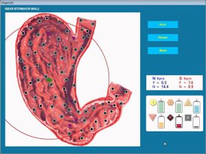
2. T2 phase: tumorous infiltrate within the first and second layers of the
stomach wall accompanied by hyperchromogenic density of the first two lay?
ers (5?6 points at the lesion spot).
3. T3 phase: a frank chromogeneity of the inner layers of the stomach
wall (6 points) except for the serous layer, which is evaluated at 4 or more
often 5 points.
4. T4 phase: lesion of all layers of the stomach wall accompanied by
chromogeneity of the serous membrane (6 points) and signs of tumorous
invasions into the neighboring anatomical structures accompanied by a frank
chromogeneity (4?5 points) of the adjacent organs.
Detection of some affected peregastric (regional) lymph nodes and dis?
tant metastases in the course of investigation allowed to analyze the N and M
criterion as well.
45
words, the NLS investigation method allows to diagnose stomach tumors
even in early phases of the disease.
Most literature dealing with the use of radiological computer tomogra?
phy to diagnose stomach tumors provides a proof that this method can poten?
tiality be used to diagnose gastric cancer, especially its endophytic forms.
However, most authors still believe that the principal role of this method lies
in acquisition of certain very important information about the extent of stom?
ach lesion and spread of the process to some adjacent organs. According to
different researchers, the early gastric cancer, that only affects the mucosa
and submucous layer can not be detected on the computer tomograms. In the
authors’ opinion, this is beyond ‘the resolution capabilities’ of this investiga?
tion method.
This work attempted to evaluate the potentials of the noninvasive radia?
tion methods of investigation (trans?abdominal ultrasound scanning and
radiological computer tomography) in detecting intramural invasion of gas?
tric cancer, and to draw their comparison characteristics.
The analyse included 72 cases of gastric cancer. All the cases were com?
pared with the surgical intervention data and the morphological studies of
post surgical evidence. According to the latest gastroenterological TNM clas?
sification of tumors (1997), the group of gastric cancer carriers in phase T1
made 9 (12.5%) cases, T2 8 (11.1%) cases, T3 22 (30.6%) cases and T4 33
(45.8%) cases. Computer nonlinear diagnostics (NLS) and radiological
computer tomography (CT) of the stomach were performed as supplemen?
tary investigation methods deliberately after a preliminary integrated
radioendoscopic investigation.
Computer tomography of the stomach was done after expanding the
stomach walls with a gas (pneumoscanning) in standard projections (lying on
the back and belly); the transabdominal NLS?investigation of the stomach
was performed using the standard procedure.
In order to more clearly comprehend the NLS and CT signs underlay?
ing the presurgical diagnosis of the T?phase of gastric cancer (i.e., invasion
degree) one needs to have a clear idea of the image of a ‘normal’ stomach wall
visualized by means of the investigation method.
Thus, in CT investigation the stomach walls (adequately expanded) were
at most 0.3 cm thick in normal conditions (test group of 50 persons) in all
regions with few exceptions in cardiac and prepiloric regions where the walls
were 0.4 cm thick, whereas at an intramural tumorous effect the stomach wall
authentically thickened over 0.6 cm (p < 0.01). In most cases it proved to be
impossible to differentiate the lamellar structure of the stomach wall by com?
puter tomography. Changing the section thickness, pitch of the table and the
44
So, an integrated approach to the use of NLS investigation and radio?
logical computer tomography has proved to be more preferable for more
accurate presurgical diagnostics of intramural invasion of gastric cancer,
however the order of priority and efficiency in their use somewhat depend on
the results of primary radioendoscopic investigation of the stomach. In addi?
tion it should be noted, that contradistinction of these methods of investiga?
tion in diagnosing and phasing of gastric cancer against each other would be
a mistake and delusion.
In conclusion, it should be emphasized that despite their subordinate
use with reference to radiological and endoscopic methods of gastric cancer
investigation, the NLS investigation and radiological computer tomography
should be brought into line with primary methods of stomach investigation.
The conclusion is based on the facts that unlike some conventional radi?
ological and endoscopic methods of investigation they allow to evaluate the
internal structure of the stomach wall, which is a master factor in the presur?
gical detection of intramural invasion.
This allows to work out the proper approach for treating patients affect?
ed by gastric cancer, and based on the well?founded data reject the explo?
rative laparatomy in case of a frank process. Considering general accessibili?
ty, lack of radiation exposure and application simplicity it appears more
apropriate to use NLS investigation as the most preferable of the above?men?
tioned methods.
47
With respect to the potentials of computer tomography in presurgical
determination of the extent of intramural invasion of gastric cancer, it should
be admitted that it had a less specific pattern and was essentially based on the
extent of the stomach wall thickening at the lesion spot.
Thus, since the CT signs are indicative of one or another degree of gas?
tric cancer invasion, they could be conditionally classified in the following
manner:
• It proved practically impossible to distinguish between tumors in T1
and T2 phases. So their diagnostics was based on the analyses of non?multi?
ple stomach wall thickenings from 0.3 to 05 cm, with the external outlines
being clear and smooth.
• The T?3 phase typically had integer multiple thickenings of the stom?
ach wall over 0.5cm not accomapanied by deformed external outlines of the
stomach wall and with no signs of the tumor spreading beyond the stomach
wall ,
• The T4?phase had multiple thickenings of the stomach wall (two,
three or more times as thick) over 0.1cm with a disturbed integrity of the
external outlines of the stomach wall at the lesion spot and with some signs
of tumorous invasion into the adjacent anatomical structures.
According to our information, the NLS?investigation proved to be the
most accurate and specific method of investigation in presurgical diagnosis of
gastric cancer in its early phases (T?1, T?2) while CT results appeared to be
more convincing in detecting later phases of tumorous lesion (T?3, T?4). It
should be noted, that, in our opinion NLS is the most accurate method of
investigation in detecting remote metastases (p > 0.05). Based on the statis?
tical analyses, the specificity of the NLS method of the investigation in
detecting the T?phase of gastric cancer (with calculations made with refer?
ence to T?1, T?1 phases) amounted to 76%, sensitivity to 74.3% and accura?
cy to 78.2%, w.r.t the computer topography the specificity, sensitivity and
accuracy were 70% each (in this case calculations were made w.r.t. T4?phase
of gastric cancer, because differentiating the lamellar structure of the stom?
ach wall was found impossible in CT investigation).
Thus, as compared to computer tomography, the NLS investigation
proved to be a more specific method for diagnosing gastric cancer in its early
phases although in a number of cases it was found difficult to differentiate
between T1?T3 phases of a tumorous lesion. In CT investigation T1?T2 phas?
es were defined conventionally based on the degree of stomach wall thicken?
ing at the lesion spot. NLS did not succeed in imaging anatomical structures
beyond the stomach wall as distinctly as CT investigation did, but NLS was
more efficient in evaluating such characteristics as M and N.
46
important, for example, for differential diagnostics. As a result of using high
frequency sensors in combination with virtual imaging equipment, a high
spatial resolution is provided which is very important for patient examina?
tion.
According to the analysis of our observations, the correct diagnosis was
made for 22 patients of 23 (95.6%). In one female patient with Itzenko?
Kusching syndrome in clinical evidence the NLS?scopy detected in the supe?
rior pole area of the right kidney a small (30?35 mm) hyperchromous growth
which had a monochromous internal structure and a spectral similarity to the
‘adrenal adenoma’ reference standard (d=0.217) and was regarded as a
benign adrenal tumor. Because of typical clinical presentation of Itzenko?
Kusching syndrome and concomitant changes in blood and urine in clinical
and lab evidence, computer tomography was not performed. The surgery
detected a focal macronodal hyperplasia of the adrenal, which was confirmed
by the histological investigation of the removed growth.
Generally a distinction is made between the diffuse and focal hyperpla?
sia. Focal hyperplasia can be subdivided into micro? and macronodal hyper?
plasia. As a matter of fact, focal hyperplasia can not be easily discriminated
from adrenal tumors by means of NLS evidence, so in this partcular case our
conclusion cannot be considered to be diagnostically wrong.
Of 23 cases computer tomography was done for 17 with its data match?
ing the NLS?investigation results. 9 patients of 23 underwent surgery to have
an adrenal tumor removed, and the NLS data were confirmed by surgery and
histological investigation results.
The size of detected tumors varied in diameter from 6 to 10 cm. All of
them had a capsule, smooth surface and rounded or oval shape. Small tumors
had a monochrome structure and in 4 cases with the tumor size over 8 cm,
the internal structure was represented by an irregular alternation of areas
having different color saturation on account of necrotic zones, degenerative
changes and decalcification, which was confirmed by a histological investi?
gation of the removed tumors.
According to the literature and our information the differential diagnos?
tication of benign and malignant adrenal tumors is an extremely difficult
process. A malignant pattern of the detected growth can be suspected only in
the presence of an irregular internal structure of the growth, restricted kidney
mobility at forced breathing, enlarged regional lymph nodes and metastases
in other organs.
Thus, diagnosis of adrenal tumors is complicated because of diverse
clinical implications in place. The potentials of the NLS?diagnostics could
scarcely be overestimated, for the simplicity of the investigation procedure,
49
Potentials of NLS?scopy
in adrenal tumor diagnostics
B.I. Lukashenko,
T.B. Georgadze
Adrenal tumors do not occur very often still they are known to be most
often hormon?active and even if small sized cause various glandular disor?
ders. However, practice knows adrenal tumors, which clinically develop with
no symptoms in evidence or are accompanied by vague complains.
Both documentary evidence and our experience show that from 6
months to two years may elapse from primary disease manifestations to form?
ing a diagnosis. Early detection of adrenal tumors has become an important
clinical problem, which is now conventionally solved by means of ultrasound
investigations (US) along with computer tomography (CT), magnetic reso?
nance tomography (MRT) and angiography (AG).
Today NLS?scopy is one of the most advanced informative hardware?
based methods of diagnostication. The NLS method will allow to substan?
tially increase the percentages of early and accurate diagnostication of adre?
nal tumors. The implementation of the latest devices equipped with digital
trigger sensors made it possible to detect any growth sized around 1 cm in
adrenals, which is comparable to computer tomography in terms of diagnos?
tical accuracy.
This our research aimed to study the NLS?scopy potentials in adrenal
tumor diagnostics and to provide an evidence that with a proper procedural
approach and advanced equipment in place, bulky growths could be diag?
nosed as successfully as if by using computer?aided and magnetic resonance
tomography.
Subject and methods
Clinical data: from June 2000 through May 2001 23 patients aged 25?64
were examined who were suspected to have developed adrenal tumors based
on careful complaint analysis and clinical and lab data. For all the patients
kidney and adrenal investigation was conducted under a regular procedure by
means of the Oberon metatron equipped with a 4.9 GHz digital sensor and
developed at the Institute of Practical Phychophysics. The device has an
automatic focusing, that can self adjust both when emitting and receiving
echo signals, and it ensures high definition of the spectrogram, which is very
48
Diagnosing rare instances of mammary gland
diseases using NLS?investigation
S.N.Okunev,
K.S. Kogan
This article deals with some clinical observations of rare cases of mam?
mary gland diseases detected by means of nonlinear diagnostics method
(NLS) during clinical diagnostic prophylactic investigations performed in
October 2001 with the purpose of an early diagnostics. In the course of the
activity the examined patients were found to have different diffuse or nodal
mammary gland diseases among which of particular interest are some rare
diseases like liposarcoma (1 case), colloidal cancer (1 case), phyllode cys?
tosarcoma (1 case), phyllode fibroadenoma (2 cases), hemangioma (1 case)
and Mondore’s syndrome (1 case).
According to some literary evidence, colloidal cancer and liposarcoma
do not occur very often among malignant tumors: colloidal cancer in 2,4%
and liposarcoma in 0.001? 0.03% of cases respectively. Among benign tumors
hemangioma occurs in 0.12%, phyllode fibroadenoma in 5.4% and phyllode
cystosarcoma in 2.5?5.4% of cases. The investigation was done using series
4011 Oberon device with a 4.9 Ghz nonlinear sensor under the program of
virtual tissular representation of panoramic scanning (Panoramic NLS
Imaging).
Mammary gland liposarcoma
Patient G., aged 28, was admitted having complaints of a nodal forma?
tion in her left mammary gland. During the examination an umbilication
symptom in the upper outside guardant of the left mammary gland was dis?
covered. The palpation detected a small?sized painless sluggish node of a
dense elastic consistency, that had a doled structure and adhered to the skin.
By means of a detailed imaging the investigation ascertained a node of 15?20
mm in diameter and detected its spectral similarity to the reference standard
‘mammary gland liposarcoma’ (D=0.204). The investigation of the regional
lymph nodes did not detect any metastatic lesion.
Based on the NLS?investigation data a surgery (radical mastectomy
according to Maden’s method) was performed. The histological diagnosis
was liposarcoma.
51
its harmlessness and rich informational content allowed us to make a correct
and promt diagnosis in 78% of cases. The conducted investigation showed
that NLS?scopy may become as efficient method of adrenal tumor examina?
tion as computer tomography and US. Patients having complaints typical for
adrenal tumors should be examined by means of the NLS?method the first.
Patients with symptoms indicative of adrenal tumor should be referred to spe?
cialized endocrine surgery departments where they would be thoroughly
investigated using US, CT and MRT which will allow to substantially reduce
the investigation time and timely provide a correct treatment.
50
were performed. The histological diagnosis was phyllode fibroadenoma.
After the plastic surgery (one month later) a repeated NLS?investigation
was carried out.
Mammary gland hemangioma
Patient C. aged 42 was admitted as having complaints of a large size
node in the right mammary gland.
During the examination it was found out that the skin over the node had
a cyanotic cast. On the border between the upper quadrants a mobile forma?
tion with a soft consistency was palpated. The graph analysis detected a sim?
ilarity to the reference standard ‘hemangioma’ (D=0.414).
The patient underwent surgery, i.e., a sectoral resection was performed.
The histological diagnosis was cavernous hemangioma.
The presented clinical observations showed that the continuous
improvement of NLS?systems and the modern technologies made it possible
to introduce a morphological picture of the neoplasm which helps devise the
optimal tactics of surgery with further morphological verification of the der?
rived results.
53
Colloidal mammary gland cancer
Patient K. aged 49 was admitted as having complaints of a node in the
left mammary gland.
The examination did not produce any visible evidence of changes in the
mammary glands. A sluggish roundish formation with a diverse consistency
and vague outlines was palpated on the border between the upper quadrants
of the left mammary gland.
The NLS?investigation detected a roundish formation (6 points accord?
ing to Flandler’s scale). The graph examination showed spectral similarity to
the reference standard ‘solid cancer’.
The patient underwent surgery; a radical mastectomy was performed
according to Moden’s technique. The histological conclusion was colloidal
cancer.
Phyllode mammary gland cystosarcoma
Patient G. aged 46 was admitted as having complaints of a slow growth
dense node in the right mammary gland.
Palpation detected in the upper outside guardant of the right mammary
gland a painless motionless formation with a diverse density and heteroge?
neous surface, that adhered to the skin.
The NLS?investigation detected an irregular 3x6x4 cm formation. A
spectral similarity to the reference standard ‘mammary gland sarcoma’
(D=0.412) was found. Its small size allowed to completely visualize the
involvement of lymph nodes.
The patient underwent surgery ? radical mastectomy according to
Moden’s techique. The histological diagnosis was phyllode cystosarcoma.
Phyllode mammary gland fibroadenoma
Patient Z. aged 43 was admitted as having complaints of a large node in
the right mammary gland.
The examination showed asymmetry of the mammary glands.
On the border between the outside quadrants of the right mammary
gland a mobile node of a large node with distinct outlines and smooth surface
and tight elastic consistency was palpated. The skin above the tumor was very
thin.
The NLS?investigation detected some hyperchromous structures corre?
sponding to intranodal vessels. The analysis of the graphs detected a similar?
ity to the reference standard ‘fibroadenoma’ (d=0.384).
On the strength of the NLS?investigation subcutaneous amputation of
the right mammary gland and omentomammoplaty on a vascular pedicle
52
with granulomatous fields which are small ? 2.0 cm or less in diameter. On
NLS image these formations look like hyperchromatic areas (6 points on
Flandler’s scale) with the central necrosis zone in ring?shaped structures
being visible as an area of lower chromogenic density (4?5 points).
Hemorrhage areas of small size are quite typical. The above?mentioned
changes are localized peri?and paraventriculary, often in the region of bor?
dering corticomedullary structures as well as in the region of basal ganglia.
Clinical observation
Patient K., born in 1974. The preliminary diagnosis when admitted into
the neurological department was acute cerebral circulation disturbance in the
spinal artery basin.
The patient complained of weakness in left limbs, speech impediment,
asthenia and loose cough. Accourding to her wording she fell ill (Feb. 06.01)
when she stopped talking, developed weakness in left limbs, diplopia and dis?
turbed swallowing. The anamnesis read a developed right?side hemiparesis,
that passed off by itself within two weeks.
The patient is in grave condition. Neurological status: conscious, under?
stands when she hears people speaking to her, but doesn’t speak. Cranial
nerves: equal palpebral fissures, nystagmus not present, the right nasolabial
fold smoothed out. Slight deviation of the tongue to the right. High tendon
and periosteal extremity reflexes, weakness in right limbs. Reduced pharyn?
geal reflex on both sides. Rigidity of occipital muscle is moderately frank.
Kering’s symptom on both sides. Babinski’s reflex on the left.
Laboratory investigation:
Clinical blood analysis: erythrocytes — 3.96×10.12/1; hemoglobin — 127
g/1; ?0.9; L — 5.6×10.9/1, ESR — 32 mm/g, — 1; — 74; — 21; — 2; — 2.
Biochemical blood test: glucose — 4.6 mm/1; urine — 6.6 mm 1/1,
bilirubin — 13.38; — 5.5; — 7.85 mmo1/1; creatinine — 0.066 mmo1/1, total
protein — 70.0m g/1; albumin — 5.5; globulins — 44.8; L2 — 4.8; L2 — 7.7,
B — 11.8; J — 20.5; — 1.24; — 0.29 mmo1/1; — 0.31 mmo1/1.
Test for toxoplasmosis detected antibodies with rising antibody titer in
dynamics (1:21 — 1:400).
Cerebrospinal — 2.5 mmo1/1; chlorides — 1.24 mmo1/1, protein —
0.2 g/1; sugar — 4.1 mmo1/1. Cellular composition: cytosis — 213, L — 1?2;
erythrocytes — 4?5.
MRT?investigation of the encephalon (of 16 Feb. 01) in T?2 picture on
both sides, detected paraventriculary and subcortically multiple unequal?
sized roundish nodi (from 0.5 to 2.0 cm) producing an unevenly increased
55
Diagnostics of toxoplasmosis (serologic
examination, CT, MRT and NLS)
T.I. Pankova,
A.S. Sosnovskaya
Toxoplasmosis is a parasitic disease whose causative agent is toxoplasma
(Tixoplasma gondii Nicolle et Manceaux), which belongs to protosa. The
disease typically has a chronic course, nervous system lesion, limphadenopa?
thy and enlarged liver and spleen. Quite often myocardium, muscles and eyes
are affected.
The infection is mostly transmitted through the alimentary tract. Yet
there are some instances recorded where contagion occurred through the
injured skin and mucous membranes. Toxoplasma is apt to form cysts in tis?
sues causing a latent infection condition. The parasite becomes active in the
conditions adverse to the macroorganism and with its immune responsive?
ness going down. In the pathogenesis of the toxoplasmodial lesion of the cen?
tral nervous system of importance are local inflammatory occurrences, dis?
circulatory disturbances related to vasculitis and blocked liquor tracts leading
to hydro? or microcephaly.
Clinically, the lesion of the central nervous system manifests itself as
meningitis, encephalitis, meningoenciphalitis and encephalomyelitis.
The most typical form of toxoplasmosis of the central nervous system is
meningoenciphalitis, which clinical picture contains general cerebral and
meningeal symptoms, paresis and limb paralyses, tonic and clonic spasms,
opticokinetic?(diplopia) and coordination disturbances. The blood test
reveals a left?shiifted leukocytosis and increased ESR; the cerebrospinal fluid
contains lymphocytic pleocytosis and a moderately increased protein con?
tent.
In diagnosing toxoplasmosis of importance are cranial radiography,
serological investigation, pneumoencephalography, CT and MRT. However,
it is the NLS?investigation of cerebral structures that plays the most impor?
tant part in the diagnosis. Of great diagnostic importance is a substantially
increased spectral similarity to the reference standard ‘toxoplasma gondii’ (D
< 0.425). The toxoplasmosis should be discriminated from viral encephalitis,
encephalomyelitis and meningitis.
In the MRT?investigations toxoplasmosis is manifested by a progressive
multifocal encephalopathy. With toxoplasmosis the typical cases have to do
54
Nonlinear diagnostics
of thyroid gland pathology
K.M. Beznogov,
L.V.Kondratyev,
S.N. Pauli
1
The spectroscopy of biophotons in non-local genetic regulation
P.P.Gariaev, G.G.Tertishny, A.M. Iarochenko, V.V.Maximenko, E.A.Leonova
Wave Genetics Inc.
Toronto, Canada
(Note: Due to considerable translation difficulties, the editors suggest that you contact the authors for verification
before quoting this material)
Abstract: This paper describes the phenomenon of broadband radiowave radiation (RR)
produced by a special optical quantum generator. It is shown that RR can be used as the basis of
polarization/lase/radiowave spectroscopy of substances. The spectroscopy mechanism closely
connected with inelastic scattering and photon localization in electronic systems of laser mirrors
is physically and mathematically formalized. This differs from the traditional Raman effect of
photons. The spectrum of inelastic scattered light is continuous and occupies the full frequency
range from 0 up to 2w (w- frequency of scattering photon). The mechanism of an EPR (Einstein-
Podolsky-Rosen) effect for localized photons is offered. It is shown that the existence of unique
localized photons (rather than EPR-correlated photon couples) is sufficient to transmit signals
instantaneously (permissive teleportation). It is shown that RR, read from DNA samples, carries
morphogenetic signals. RR of DNA induces in recipient plants morphogenetic modifications and
is also capable of repairing radiation-induced genetic damage in plants. It has been proposed
that RR transmission from DNA to recipient plants takes place through permissive teleportation.
I. The general working principles of a laser installation showing the phenomenon of
transition of optical photons to radiowaves.
Previously we developed a laser installation with the help of which we have found the
phenomenon of transition of red coherent photons to radiowaves of a wide spectrum. We have
offered a preliminary explanation of this phenomenon [21]. The present research offered by the
authors essentially supplements the positions earlier stated by them and is at the stage of a
theoretical-experimental substantiation of a new kind of spectroscopy of substances with the
conditional name “polarizing laser-radio frequency spectroscopy” (PLR-spectroscopy). Such
spectroscopy is intended for research into previously unknown, rotational-vibrational quantum-
molecular characteristics of solid, liquid, gaseous substances, and also plasma states. The variant
of PLR-spectroscopy offered uses a narrow optical range red light, but further developments are
being planned which will make use of more short-wave spectra in the visible region.
For the purposes of PLR-spectroscopy a special He-Ne laser (632.8 nm)was used to generate
two orthogonal optical modes correlated in intensity in such a manner that the sum of their
2
intensities remains constant. Upon interaction of one mode with the target substance, the
reflected, or non-local, radiation is returned to the optical resonator, [where] thereis a
redistribution of intensity of these optical modes, under the law of change of polarization
appropriate to a new condition after interaction of a beam with dynamic micropolarizers, which
takes place in a cross-section of an illuminated platform of the target substance. One of the laser
modes, at a certain mode of generation, is able during interaction with the target substance to
cause radiation by our installation of modulated radiowaves of a wide spectrum, correlated with
modulations in optical modes of radiation of the laser. These modulations depend on rotary
fluctuations of microstructural components (for example, domains of crystals) of the target
substances and their optical activity.
The frequency interval of the induced radiowaves, according to the theoretical model (see
below), lies in a range from 2 up to 0. The maximum of such radio emission settles down in the
1 MHz region. The radiowave signal after detection is transferred to an analogue numeral
transformer on a computer with a special processing program. On a display is registered the
Fourier spectrum of a radio emission describing polarization-dynamic properties of the
investigated substances with which one of the laser beams interacts, and also the spectral
memory of investigated substances. The second beam thus comes back to the laser resonator for
creation of resonant interaction with atomic oscillators of the gas mix. The given laser also is
able to generate, except for the basic (optical) frequency, a radiowave ofa wide range of wave
lengths. The reason for this phenomenon is, we believe, the inelastic scattering and localization
of light of the basic laser mode on system heterogeneities in the mirrors of the laser resonator.
The mechanism of localization (localization in the inelastic channel of dispersion) is described in
detail. In particular, the position is put forward, that in the resonator there exists as well a form of
elastic, non-local light (see theoretical part).
Radio frequency radiation generated by the laser is able “toread outthe information”, for
example, from DNA preparations (see experimental part). The mechanism of “reading” is similar
to the mechanism of usual induced radiation. The opportunity “to open and close” thelaser
resonator makes it possible “to locate or write down”in itself “spectra” of various tested objects.
Radio frequency radiation reads out and relays such spectra. Thus the effect of spectral memory
was identified: for a certain macroscopic time, radio frequency spectra of the objects reflecting a
beam back into the resonator and then removed from the examination zone, continue to be
reproduced. So DNA spectra were registered and their high biological activity, probably
connected with wave-type transmission of genetic-metabolic information, was revealed (see
experimental part).
Experimentalpart
II. PLR-spectroscopy of minerals and biostructures. Effect of spectral memory.
3
In Fig. 1 the circuit of typical experiment with the record of a PLR-spectrum of target substances
(for example, mineral crystals), is shown.
Fig1. The experimental circuit with arecord of a PLR spectrum
Fig2
Fig. 2 the PLR-spectrum of a mineral (apofillit). Arrows (pointers) specify area of display of the spectrum,
given inFig. 2a.
4
Fig2a
5
6
Fig. 2a. Polarization-Laser-Radiowavedisplayof a spectrum of a mineral apofillit
Frequency of digitization of a signal 44 kHz. Areas 1550-1660 Hz, 1660-1760 Hz, 1760-1860
Hz are developed (unwrapped). It is clear that these areas of the spectrum have isomorphic
structure with differing amplitudes. Such type of spectral modulation can be named
heterogeneous frequency fractalization modulation.
Fig 3: record of a PLR-spectrum (frequency of digitization of a signal – 22 kHz) of a live green [leaf?] of a
wheat seedand spectral memoryof this object.
Before the experiment, as in the case of the turmalin and apofillit crystals, we fixed a background
radio emission of a PLR-spectrometer which was typically noise, and its amplitude was
exponentially reduced to 5000 Hz. For live leaves the characteristic expressed frequency areas
were identified as 800-900 Hz, 1700-1900 Hz, 2400-2600 Hz and 3600-3800Г Hz . After
removal of the wheat seed the PLR-spectrometer continues for some time to generate a radio
7
emission, characteristic for wheat leaves. In it spectral PLR-memory is also shown.
Fig4
Fig 4: PLR-spectra of highpolymerization DNA sample from calf thymus (the top spectrum) and its spectral
“trace” on lasermirrors (the bottom spectrum) after removal of a DNA sample from a zone of probing laser
beam. As in the case of minerals and wheat seed,the affinity of a spectrum of preparation DNA and a
spectrum of its “trace” is visible.
III. Biological activity of PLR-spectra of DNA samples
“Recording” thePLR spectra ofDNA samples proved to have a specific effect on the biosystems
we used – for example, inducing abnormally fast germination (up to 1cm/day) in a potato plant,
or revitalizing seeds of the plant Arabidopsis thaliana which had been damaged by radiation
8
from the Chernobil Atomic Power Station accident in 1986-1987. In a typical experiment
looking at the effect of three different PLR protocols on such seeds, (1h. 30min., 1h. 40min. and
2h. treatments with a radiation dose of 25 mR/hour) the “DNA-radiowave-radiation” in last two
groups was observed to significantly increase the germinating capacity of seeds in comparison
with two control experiments (P <0.001). That is, from 300 and 200 sown seedsin thecontrol
only 2, respectively 4 seeds germinated – while in the experimental group 16 and respectively 24
seeds germinated. However, after the dose was increased to 170 mR/hour the effect of “seed
revival” appeared to reach a plateau.
This shows that the radiowave DNA emission obtained in this way has the ability to restore the
genetic control apparatus and vitality of A. Thaliana seeds, but within limited intervals of
radiation dose capacity. Essentially the seeds were stored for a long time (1987 -1999), and that
has resulted in their significant ageing, imposing an additional damage factor. Nevertheless, a
“revitalization” effect is observed, and it demonstrates that DNA- radiowave radiation can carry
in itself reparative genetic (metabolic) information that confirms our early work on wave biosign
reparative influences on X-Ray irradiated wheat and barley seeds [17, 18]. It is likely that the
recognition of such wave information is carried out by seed – acceptors on the level of a quantum
nonlocality (teleportation) mechanism, as we assumed earlier [5, 19], but here we intend to
update the permissive model offered in the previous research.
Before discussing the theoretical-physical analysis of the offered teleportation model (see
below), we will state some opinions concerning the importance of this problem for genetics and
biology as a whole.
In works [5, 19] the question of genome information quantum teleportation was already
discussed. In the present research these ideas are formalized and consequently aremore
thoroughly supported. Presumably it is possible to interpret the biological experiments
mentioned above as a demonstration of the imprinting of genetic information from DNA
preparations on biosystem-recipients through the mechanism quantum teleportation in
permissive variant. It is proposed that the quantum nonlocality of the genetic (chromosomal)
information as displayed in its total continuity in the space of multicellular biosystems, is a
special case. Actually, in biosystems, at least, there are six levels nonlocality:
1st level: Level of the organism. Nonlocality here is expressed in the capacity for regeneration,
for example of planarium worms. After cutting these worms, any part of their body regenerates
into the whole organism. Differently stated, in this case there is no binding of the genetic
information to any part of the biosystem. The same concerns to vegetative duplication of plants.
2nd level: Cellular. From each cell, and not just from a zygote, it is possible to express the whole
organism. For animal biosystems it is complicated, but it is possible. Each cell – a potential
continuum of an organism.
3rd level: Cellular – nuclear. Enucleating a nucleus from a somatic or sexual cell, with the
subsequent introduction into this cell of another nucleus, does not interfere with the development
of a normal organism. Such cloning has already been carried out on higher biosystems, for
9
example, on sheep. Each cellular nucleus is also a potential continuum of the biosystem.
Localizations of genetic potentialities on any separate cells are not present.
4th level: Molecular. The ribosome “reads” and interprets the messenger RNA not only through
separate codons, but also globally, dependent on a non-local “context”.
5th level: Chromosomal/holographic. A gene has holographic memory [26], and it is typically
distributed (non-local) associative memory. On this and the subsequent levels nonlocality
receives a new quality, a dualistic matter/wave nature – as chromosomal material texts “are read”
by the electromagnetic and/or acoustic fields bearing genetic/wave information. For example,
the physical field acts as calibrating factor, creating the future space-time developmentof a
potential organism with the help of the holographic memory of the brain cortex – specifying
mental, semantic and figurative spaces, calibrating potential actions of the higher (conscious)
biosystems. Through this socio-genetic processes are realized.
6th level: Quantum nonlocality of genome. Up to the 6-th level the nonlocality of the genetic
information is realized within the space of an organism. The 6-th level has a special character
and a new quality. It is shown within the framework of one of the forms of quantum nonlocality,
namely permissive, postulated in the above-mentioned work.
In this case nonlocality is realized both in the space of the biosystem, and on its own,
“compressed-to-zero”,time. Genetic/Waveprograms instantly distributedin such ways,
(isomorphic material), work in an organism “here and there simultaneously ” [17, 18]. This is a
strategic factor of extraordinary importance for multicellular biosystems’ evolutionary
achievement. Billions of cells in an organism should “know” about each other – communicating
instantly about their status. Without the phenomenon of “wave information instantaneousness ”
the huge multicellular continuum of higher biosystems is not capable to completely coordinate
the metabolic, physiological and other functions. Intercellular diffusion of messenger substances
and nervous processes are too inert for this purpose. Even if we admit that sign electromagnetic
fields participate in intercellular transfer with light speed, that is still insufficient. The
mechanism of quantum nonlocality is necessary, and it is applicable to the genetic device which
can act as instantly distributed quantum (wave) object, isomorphic to material chromosomes [17,
18,
We are MAIKONG 3d nls health analyzer | 3d nls health analyzer price | Metatron 4025 Hunter | original 3d DIACOM nls|www.3d-nls-health-analyzer.com,manufacturers Unified Wholesale price.Welcome to inquiry and OEM.
Related Items